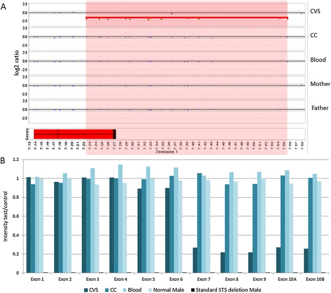
Figure 1.
(A) Array CGH results. Magnification of the STS gene and copy number loss on the X chromosome at Xp22.31. The deletion region is highlighted in red. The STS gene is shown as a red block at the bottom left. Exons are shown as black lines within the STS gene box. A copy number loss disrupting the STS gene is identified in the direct chorionic villus sample (CVS). This loss included exons 8, 9 and 10 of the STS gene. Exon 7 is located adjacent to the call, but before the next normal probe, thus it could also be deleted. The results from DNA from CVS-CC (CC), neonatal (Blood), maternal (Mother) and paternal (Father) blood samples indicate no evidence of this copy number loss. Therefore, the deletion is de novo and confined to the placenta. (B) Multiplex Ligation-dependent Probe Amplification results for the STS locus. Only proband direct CVS (CVS), CVS-CC (CC) and neonatal blood (Blood) are shown here and are compared with a normal male sample and a sample with a known standard STS deletion. An intensity ratio between 0.8 and 1.2 corresponds to a normal copy number (i.e. 1 copy in males). Males carrying the deletion will show no amplification and an intensity ratio of 0 (0 copies). It is clear that exons 7–10 of the STS gene are deleted in a mosaic state in the direct CVS sample

