Figure 2.

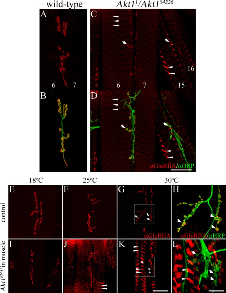
GluRIIA localization was modified in Akt1 mutants and animals with muscle-specific inhibition of Akt1. GluRIIA localization was examined in muscles 6 and 7 using monoclonal anti-GluRIIA antibody (red). Anti-HRP antibody detected neuronal projections (green). A and B: In wild-type animals, GluRIIA was located in the postsynaptic specialization that surrounds the motoneuron boutons. C and D: Akt11/Akt104226 mutants showed reduction of GluRIIA at synaptic boutons (see arrows) and redirection to intracellular bands (faint staining in muscles 6 and 7, and more prominent in muscles 15 and 16; see arrowheads). E–L: Akt1 function was compromised by muscle-specific expression of an Akt1RNAi construct using the GAL4-UAS system. UAS-Akt1RNAi/+ animals served as controls. GAL4 transcriptional activation shows temperature dependence, permitting a graded level of Akt1 blockade from 18°C (low level of inhibition) to 30°C (high level of inhibition). E–H: In control larvae, GluRIIA immunoreactivity was concentrated in the postsynaptic region surrounding boutons at all temperatures. H: Enlarged view of white box area in (G), arrows show the motoneuron boutons surrounded by GluRIIA. I–L: In Akt1RNAi expressing larval muscle (24B-GAL4>UAS-Akt1RNAi), GluRIIA mislocalization (arrowheads) was more severe with greater inhibition of Akt1 function at increasing temperature (larvae reared at 18°C (I), 25°C (J), or 30°C (K and L)). L Enlarged view of white box area in (K), arrows show synaptic boutons lacking GluRIIA immunoreactivity; arrowheads mark ectopic GluRIIA within intracellular bands. Scale bar in (A–G) and (I–K), 50 µm, in (H) and (L), 5 µm.
