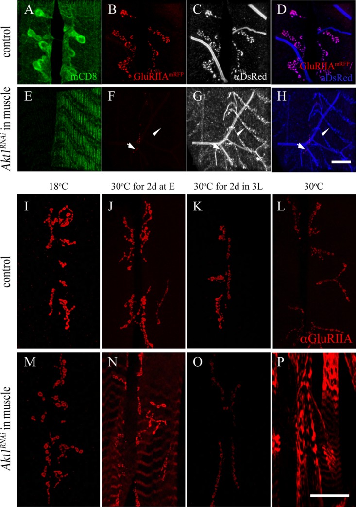
Figure 3.

Akt1 affects GluRIIA trafficking to NMJ and is crucial in the early developmental stage. Two experiments are shown here. The first (panels A–H) shows the results from a study where the distribution of an engineered GluRIIA-RFP when Akt1 function was compromised with RNA interference. The GluRIIA-RFP was detected with either endogenous fluorescence from the mRFP or an anti-mRFP antibody (anti-DsRed). The second experiment (panels I–P) was designed to determine the developmental window when Akt1 activity was critical for GluRIIA localization. Time-limited inhibition of Akt1 function was achieved using Akt1RNAi and a temperature-sensitive GAL80 (see “Materials and Methods”). A–D: GluRIIA-mRFP (red) was colocalized with anti-DsRed signals (gray) at the NMJ in control animals (G14-GAL4, UAS-mCD8-GFP; UAS-GluRIIA-mRFP/+). E–H: GluRIIA-mRFP fluorescence was reduced significantly at the postsynaptic density (arrows) upon inhibition of Akt1 function. The redistribution of GluRIIA-mRFP protein into an intracellular compartment was detected only with anti-DsRed immunostaining in Akt1 compromised animals (G14-GAL4, UAS-mCD8-GFP; UAS-GluRIIA-mRFP>UAS-Akt1RNAi) (arrowheads). I–P: To investigate the critical periods when Akt1 is required for GluRIIA localization at the NMJ during development, the temperature-sensitive GAL80 repressor under tubulin promoter, Tubp-GAL80ts, was used along with the GAL4-UAS binary system to allow temporal spatial regulation of Akt1RNAi expression (Tubp-GAL80ts, Mef2-GAL4>UAS-Akt1RNAi). I–L: At all temperatures, control (Tubp-GAL80ts, Mef2-GAL4/+) animals showed normal GluRIIA distribution at the NMJ. M: In Tubp-GAL80ts, Mef2-GAL4/UAS-Akt1RNAi animals at the permissive temperature (18°C), when expression of Akt1RNAi is minimal on account of GAL80ts blockade of transcription, the animals displayed a normal GluRIIA distribution. N: Incubation at the restrictive temperature (30°C) for 2 days right after egg laying induced modest GluRIIA mislocalization in muscles while much of the GluRIIA remained at the NMJ. O: Temperature shift from 18°C to 30°C for 2 days at the third instar larval stage produced reduced levels of GluRIIA immunoreactivity at the NMJ but no abnormal localization. P: Animals reared at 30°C throughout the entire developmental stages displayed severe GluRIIA mislocalization. Scale bar in (A–H), 10 µm, in (I–P), 50 µm.
