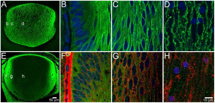
Figure 3. AQP0 and AQP5 protein distribution in the embryonic lens.

At developmental stage E16, AQP0 is expressed exclusively in fibre cells in all regions of the lens, but is not detected in the anterior epithelium (A). Furthermore, it is localised to the cell membrane of DF cells in the periphery (B), cortex (C) and PF cells in the nucleus (D). In contrast, AQP5 is localised to the cytoplasm of lens epithelial cells and fibre cells in all regions of the lens (E). This distribution continues in DF cells in the periphery (F), cortex (G), and PF cells in the nucleus (H), where cell membranes have been labelled with WGA (red) for clarity. AQP proteins were labelled with specific antibodies (green) and cell nuclei were labelled with DAPI (blue).
