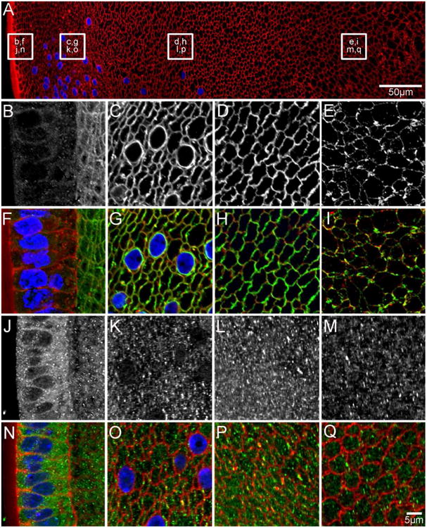
Figure 5. AQP0 and AQP5 protein distribution in early postnatal (P6) lenses.

An overview of an equatorial section from a P6 lens labelled with the membrane marker WGA (A, red) to indicate where higher magnification images for AQP0 (B-E in greyscale, F-I in green) and AQP5 (J-M in greyscale, N-Q in green) were obtained. AQP0 localises to the fibre cell membrane in all lens regions. In the nucleus, using an antibody to the AQP0 C-terminus (E/I) the initial truncation of the AQP0 C-terminus is evident due to the lower signal intensity observed in the region. In contrast, AQP5 is predominantly cytoplasmic in all lens regions, although some labelling associated with the membranes of DF cells in the lens cortex is starting to become evident (K/L, O/P). Cell nuclei (blue) are labelled with DAPI.
