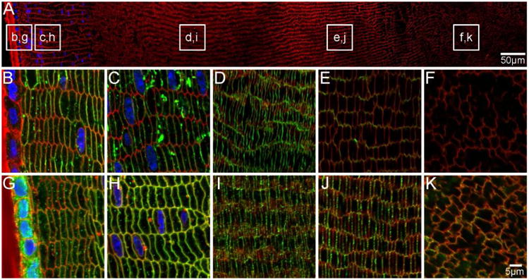
Figure 9. AQPs are localised to fibre cell membranes in aging mouse lenses.

An overview of an equatorial section of an 8 month-old mouse lens labelled with WGA (red, cell membranes) and DAPI (blue, nuclei) indicates where higher magnification images of AQP0 (B-F, green) and AQP5 (G-K, green) are taken from. AQP0 is localised to fibre cell membranes in peripheral and cortical lens fibres. Weaker signal in the inner cortex may indicate truncation of the AQP0 C-terminus (E). In the nucleus AQP0 is truncated, as indicated by a lack of signal (F). In contrast, AQP5 is localised to the fibre cell membranes throughout the lens (G-K). Some cytoplasmic labelling is maintained in epithelial and peripheral lens fibres (G).
