Figure 4.
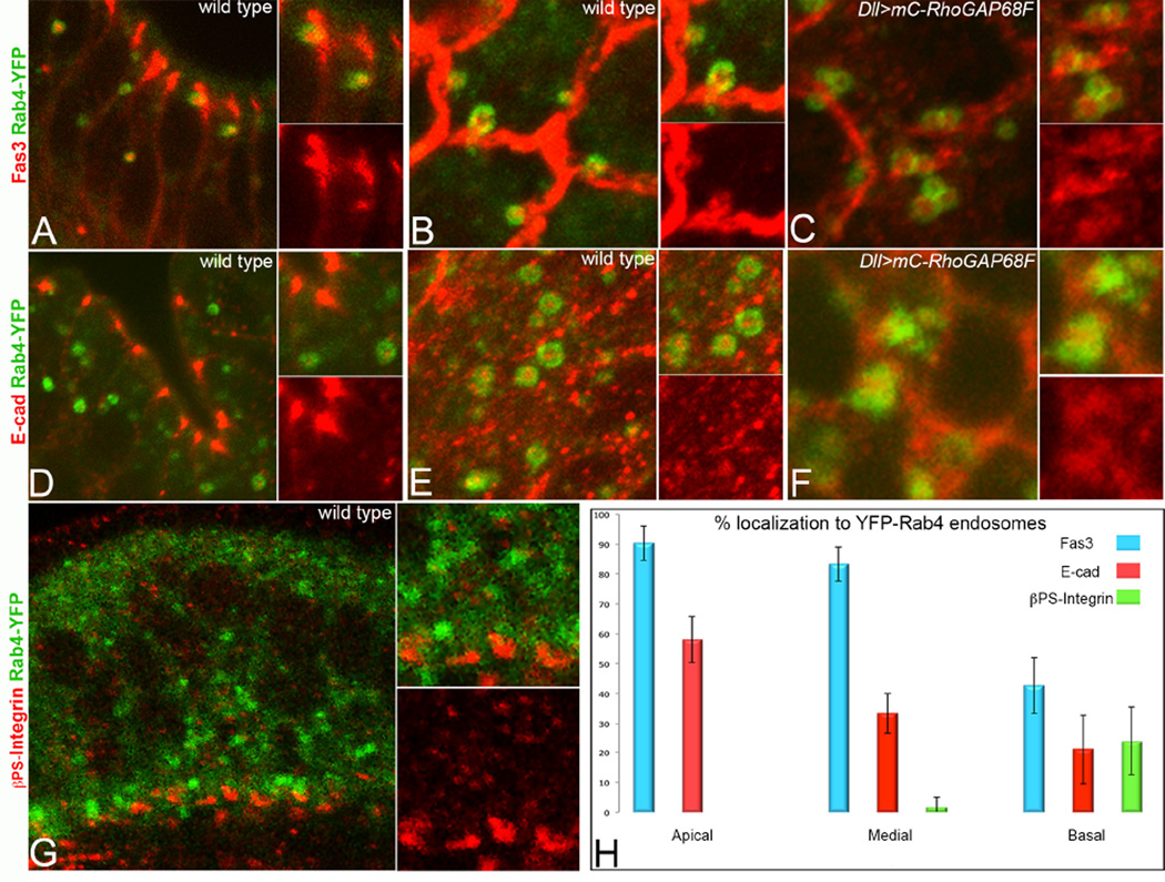
The Rab4 endosomes return the adhesion receptors Fas3 and E-cad back to the cell surface. (A, D, G) Apicobasal and (B–C, E–F) planar sections across the tarsal region at 4h APF. (A–C) Fas3 and (D–F) E-cad accumulated at high level in Rab4 endosomes and Fas3 and E-cad were clearly seen in Rab4 endosomes in the process of fusing with the cell surface (B and data not shown). Overexpression of RhoGAP68F caused clustering of Rab4 endosomes containing the adhesion proteins (C) Fas3 and (F) E-cad. (G) βPS-Integrin accumulated at lower levels in Rab4 endosomes in the basal region of the epithelium. (H) Quantification of the relative accumulation of Fas3, E-cad and pPS-Integrin in the apical (above nuclei), medial (at the plane of nuclei) and basal (below nuclei) regions of the epithelium, respectively, at 4h APF. N=3 per each adhesion protein analyzed.

