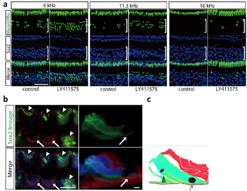
Fig. 4. Regenerated hair cells.

Hair cells and supporting cells were labeled by anti myosin VIIa (green) and anti Sox2 (blue) antibodies, respectively (a). hair cells number increased by approximately the same amount as the supporting cells number decreased. Supporting cell lineage was traced in green and other cells in red in Sox2-CreER; mT/mG double transgenic mice. (b). As described in Fig. 2, hair cells (marked by arrowheads) transdifferentiated from supporting cells were green, while surviving hair cells after noise exposure were red. Some of the green hair cells (right panel in the Z-stack) shown in the diagram (c) extended to the basilar membrane (arrow). The figure is reproduced with permission from Figure 4 and 5, Mizutari et al., Neuron Vol. 77 Issue 1.
