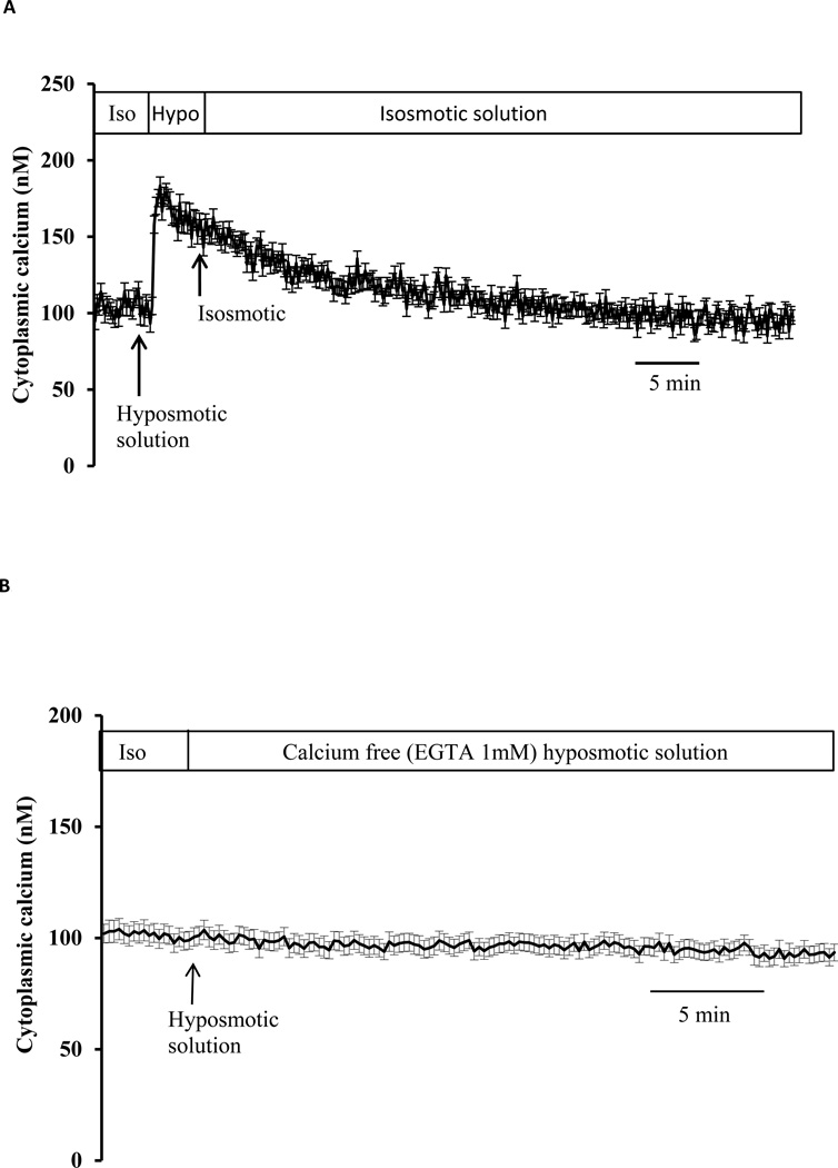
Fig. 2.
To examine reversal, cytoplasmic calcium concentration was measured in cells exposed to hyposmotic solution (200 mOsm) for 2 min then returned to control (isosmotic) solution (Panel A). To examine dependence on extracellular calcium cytoplasmic calcium concentration was measured in cells exposed to calcium-free hyposmotic solution (200 mOsm) (Panel B). Baseline calcium concentration was first measured for 5 min in control isosmotic solution (Iso) then calcium-free hyposmotic solution (200 mOsm) was introduced and the measurement continued for a further 30 min. Data from 15–30 individual cells were averaged and considered as n=1. Results are means ± SE of 5 independent experiments.

