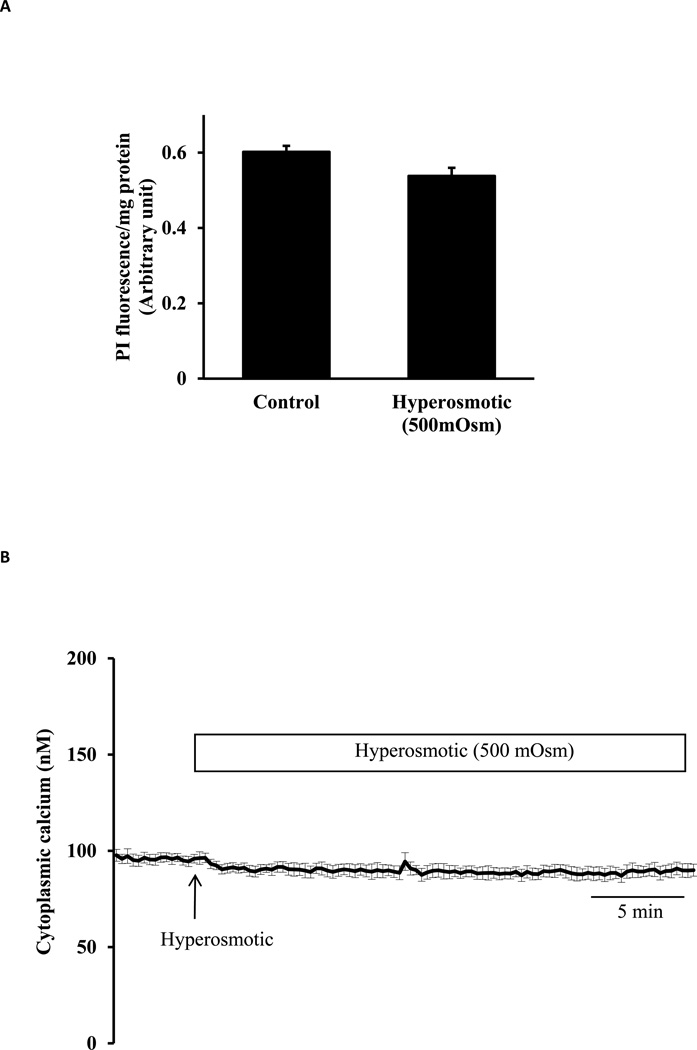
Fig. 6.
Studies on lens epithelial cells exposed to hyperosmotic solution (500 mOsm). Panel A shows propidium iodide (PI) uptake measured in cells incubated for 30 min in hyperosmotic (500 mOsm) solution (Hyper) or control isosmotic solution (Control) that contained PI (25 µM). The results are expressed as relative fluorescence/mg protein. The values are mean ± SE of results from 6 independent experiments. Panel B shows cytoplasmic calcium concentration measured for 5 min in isosmotic solution (Iso) remained unchanged when hyperosmotic solution (500 mOsm) was introduced for a further 30 min. Data from 15–30 individual cells were averaged and considered as n=1. Results are means ± SE of 5 independent experiments.

