
Figure 2.
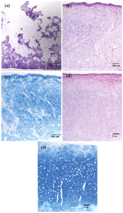
Histological sections stained with H&E and with Alcian Blue for detection of GAG. (a) H&E staining for pre-implant construct sectioned at 40μm. (b) H&E stain of post-30-day implant sectioned at 5μm. (c) Alcian Blue stain of post-30-day implant sectioned at 5μm. (d) H&E stain of post-60-day implant sectioned at 5μm. (e) Alcian Blue stain of post-60-day implant sectioned at 5μm. H&E stain of the neocartilage 30 days post-implantation shows homogeneous distribution of chondrocytes within a cell matrix. At 60 days, the implant exhibits architecture more similar to native septal cartilage.13 A moderate to substantial amount of GAG is observed with Alcian Blue staining at 30 and 60 days.