Figure 2.

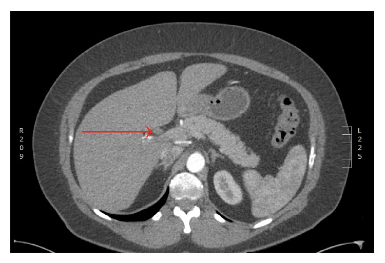
Contrast-enhanced computed tomography (CT) scan demonstrating an arterially enhancing filling defect within the proximal hepatic duct (marked with red arrow).

Contrast-enhanced computed tomography (CT) scan demonstrating an arterially enhancing filling defect within the proximal hepatic duct (marked with red arrow).