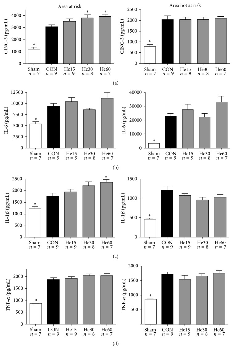
Figure 2.
Protein levels of CINC-3 (a), IL-6 (b), IL-1β (c), and TNF-α (d). Protein levels are determined in myocardial tissue after completion of the experimental protocol: all animals underwent 15 min of stabilization, 25 min of ischemia, and 120 min of reperfusion, except for Sham animals. Sham animals were not exposed to ischemia and reperfusion. Animals receiving helium postconditioning received 15, 30 or 60 min of helium. All data are shown as mean ± S.E.M. Amount of experiments in each group is shown below individual bars. Groups were tested with one-way ANOVA plus a Dunnet post hoc test comparing the control group against all other groups; * P < 0.05, significant in comparison to CON.

