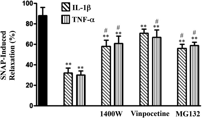
Fig. 8.
Suppression of SNAP-induced relaxation by proinflammatory cytokines and reversal of inhibition by blockade of iNOS, NF-κB, or PDE1 activity. Longitudinal muscle cells were isolated from muscle strips cultured in the presence of IL-1β (10 ng/ml) or TNF-α (1 nM) for 48 hours. In some experiments, muscle strips were cultured with IL-1β or TNF-α in the presence of 1400W (10 µM), vinpocetine (50 µM), or MG132 (10 µM) for 48 hours. Cells were treated with 10 µM SNAP for 5 minutes followed by carbachol for 30 seconds to measure initial Ca2+-dependent contraction. Smooth muscle cell contraction was measured by scanning micrometry, and relaxation was expressed as the percent inhibition of carbachol-induced contraction. Values are the means ± S.E.M. of five to six experiments. **Significant inhibition in SNAP-induced relaxation by IL-1β or TNF-α compared with control SNAP-induced relaxation (solid bar). #P < 0.05, significant reversal of inhibition by 1400W, vinpocetine, or MG132.

