. 2015 Feb;48(2):115–120. doi: 10.5483/BMBRep.2015.48.2.035

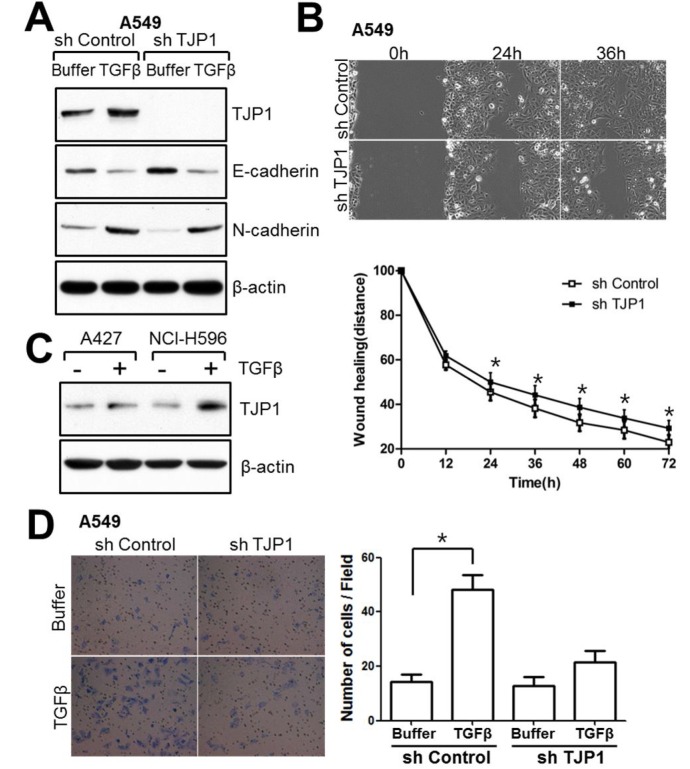
Fig. 4. TJP1 is increased in response to TGF-β in lung cancer cells and contributes to cellular motility. (A) Subconfluent cells were treated with 50 pM TGF-β for 24 h and harvested for immunoblotting. (B) Cells were grown until fully confluent, and a uniform scratch was made down the center of the well. Digital images of the wound were obtained in 12-h intervals until 72 h (top), and the horizontal distance between both sides of the wound was measured and analyzed statistically (bottom). All results shown are representative of at least three independent experiments. Data are expressed as the means ± SE. Statistical significance was assessed using paired Student’s t-tests (*P < 0.0001). (C) A427 and NCI-H596 lung cancer cells were treated with 100 pM TGF-β for 24 h and evaluated for TJP1 expression by immunoblotting. All results shown are representative of at least three independent experiments. (D) Cells were induced to invade through Matrigel- coated Transwell membranes. After 24 h, invaded cells were fixed and stained. Digital images were taken for cell counting. All results shown are representative of at least three independent experiments. Data are expressed as the means ± SE. Statistical significance was assessed using paired Student’s t-tests (P < 0.0001).

Fig. 4.