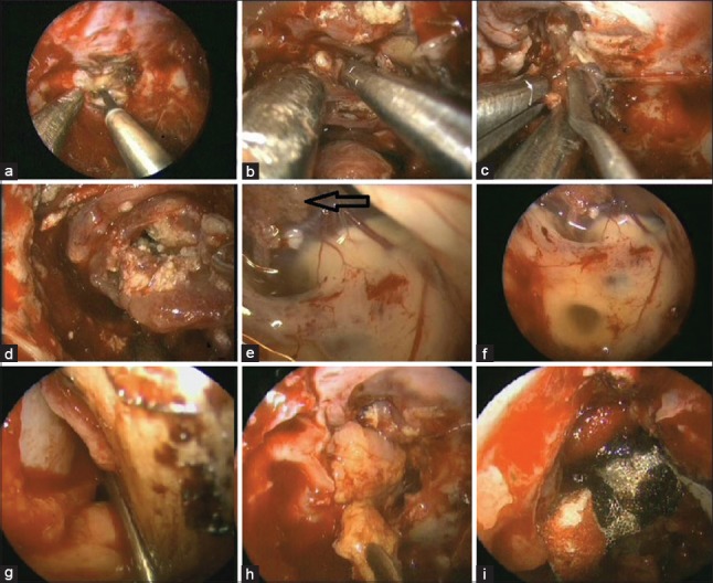
Figure 4.

Endoscopic images showing (a) coagulation of sellar and part of anterior cranial fossa duramatter, (b-d) dissection of craniopharyngioma, (e and f) visualized third ventricle with choroid plexus (arrow), (g and h) placement of vascularized flap in dural defect, (i) surgicel and tissue glue being used
