
Figure 1.
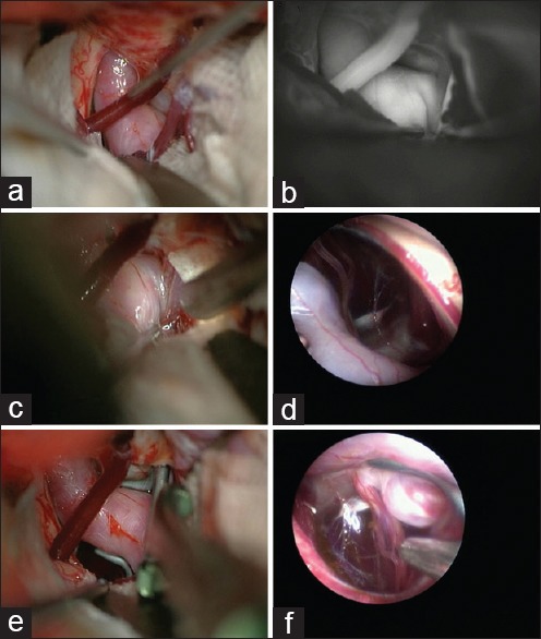
An internal carotid artery (ICA) postcommunicating segment aneurysm with the neck medially located. (a) Despite gentle retraction of the brain, the aneurysm neck and the surrounding vessels cannot be visualized well. (b) Some slight manipulation of the ICA with a suction tip to open a corridor is practiced but still indocyanine green angiography is not able to show the perforating arteries. (c) An endoscope is introduced to the field under microscopic vigilance. (d) Neck of the aneurysm as well as the medially located perforators come into the endoscopic view. (e) An angled fenestrated clip is inserted in parallel to the ICA to obstruct the medially located neck. (f) Endosopic view after insertion of the aneurysm clip to confirm complete obliteration of the neck without engulfment of the perforators