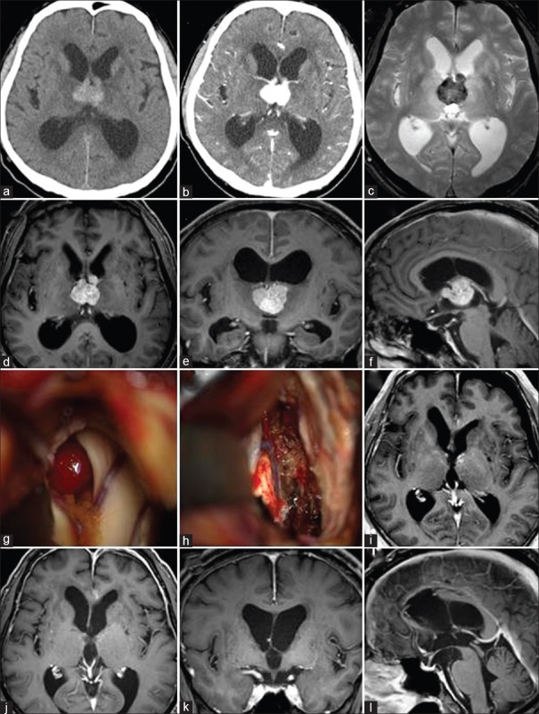
Figure 1.
A computed tomography scan and the enhanced T1-weighted image showed an abnormal mass in the third ventricle and ventricular dilatation (a, b, d-f). The T2-weighted star image showed the lesion as a hypo-intense mass that indicated the intratumoral hemorrhage (c) surgical tumor removal was performed via an interhemispheric trans-callosal trans-choroidal approach (g) gross total removal was successfully accomplished (h) a postoperative enhanced magnetic resonance imaging showed improvement of the hydrocephalus and total removal of the tumor (i) as of 2 years after surgery, follow-up enhanced magnetic resonance imaging showed no recurrence (j-l)

