
FIG 2.
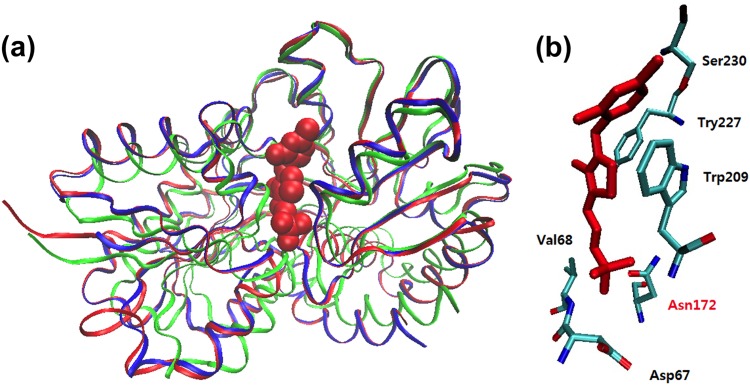
Structural modeling of TP_0144. (a) Comparison of overall structures of TDE_0143 (blue), TP_0144 (red), and the TbpA protein of E. coli (green). The TbpA protein of E. coli (PDB ID 2QRY) (36) was used as the template in structural modeling. The TPP ligand is shown in a Van der Waals representation. (b) A putative thiamine-binding site is displayed with predicted binding residues and the bound TPP (red).