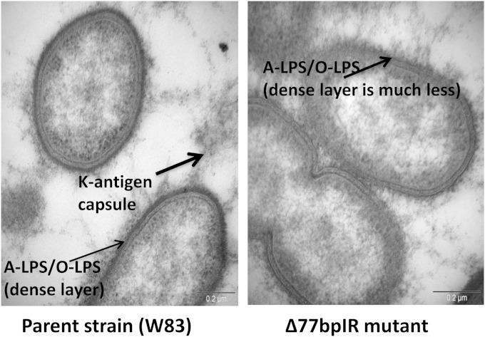
FIG 2.
Transmission electron micrographs of the parent strain W83 and the corresponding Δ77bpIR mutant. The cells were stained with ruthenium red to detect surface polysaccharides, as described in Materials and Methods. The arrows indicate the different types of polysaccharides (LPS or K-antigen capsule) on the surface of the cells. The mutant strain shows a significant decrease in the heavily stained dense layer of polysaccharide (A-LPS or O-LPS) that surrounds the parent strain.

