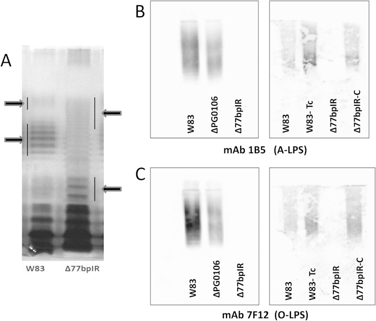
FIG 4.
LPS laddering pattern on silver-stained gels and quantification of A-LPS and O-LPS by immunoblotting. (A) Proteinase K-digested bacteria from the parent strain W83 and the Δ77bpIR mutant were separated by SDS-PAGE and stained with ammoniacal silver to visualize LPS. The Δ77bpIR mutant displayed an altered banding pattern with an overall shift to lower molecular weights. (B and C) Autoclaved extracts of W83, Δ77bpIR, and ΔPG0106 were subjected to SDS-PAGE and Western blotting, as described in Materials and Methods. Membranes were probed with antibodies reactive to A-LPS and O-LPS. The ΔPG0106 mutant has reduced reactivity to antibodies to A-LPS and O-LPS. The Δ77bpIR mutant did not react to antibody to A-LPS or O-LPS. Reactivity to both A-LPS and O-LPS was restored to wild-type levels in the complemented strain Δ77bpIR-C.

