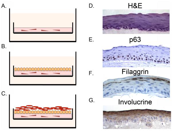
Figure 4.
Organotypic culture (OTC) technique to model human esophageal epithelium and Barrett’s esophagus in an in vitro system. A. Culture is initiated by plating human embryonic fibroblasts in a collagen matrix for 4 days. B. Human esophageal keratinocytes or Barrett’s epithelial cells are then plated on top of the collagen matrix. C. Once eithelial cells are confluent, the level of the cell culture media is reduced to place the epithelium at the air-liquid interface. D. Example of normal appearing multi-layered human esophageal epithelium that forms after EPC2-hTERT cells are grown under OTC conditions. H&E: stained by hematoxylin and eosin. E. p63 immunohistochemistry staining of an OTC culture. Immunohistochemistry for differentiation markers F. Filaggrin and G. Involucrin. Images provided by Jianping Kong MD PhD University of Pennsylvania Perelman School of Medicine.

