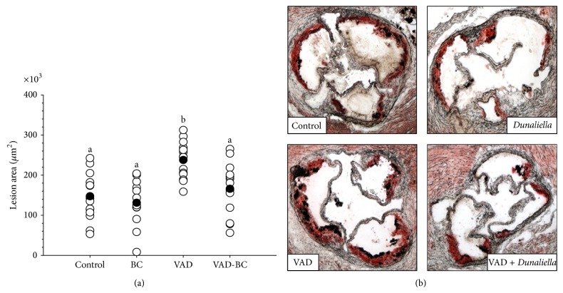
Figure 4.
Atherosclerotic lesion area in apoE−/− mice fed chow or vitamin A-deficient diet with or without β-carotene. The atherosclerotic lesion area was quantified at the aortic sinus by oil-red O staining of the lipids after 15 weeks of treatment (a). One representative aortic sinus lesion section is shown for each treatment group (magnification ×40). Red color indicates the presence of atherosclerotic lesions (b). The vitamin A-deficient diet significantly increased the atherosclerotic lesion area while β-carotene reverts this effect. Values are means ± SE, n = 13–15. a, bWithin the graph, means without a common letter differ, P < 0.05. Dunaliella treated group (BC); vitamin A-deficient diet group (VAD); vitamin A-deficient diet fortified with Dunaliella group (VAD-BC).

