Figure 5.
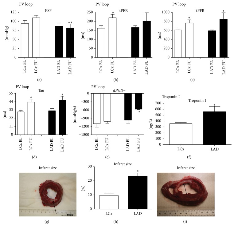
Pressure-volume loop analysis, troponin I, and infarcts size. (a)–(e) Pressure-volume loop analysis. (a) End-systolic pressure (ESP) is significantly lower at follow-up in the LAD group; (b) time to peak ejection rate (tPER); (c) time to peak filling rate (tPFR); (d) Tau: relaxation constant; (e) increase in pressure over time (dP/dt+); (f) troponin I levels 6 hours after myocardial infarction are higher in the LAD group. (g)–(i) Infarct size. (g) Example of infarct by left circumflex artery infarct (LCx) ischemia-reperfusion model. Viable myocardium is red; infarct is depicted in white (arrow). (h) Infarct size in left anterior descending artery (LAD) group is significantly higher; (i) example of an LAD infarct. LCx indicates left circumflex artery. LAD: left descending anterior artery; BL: baseline; FU: follow-up. * P < 0.05.

