Figure 3.
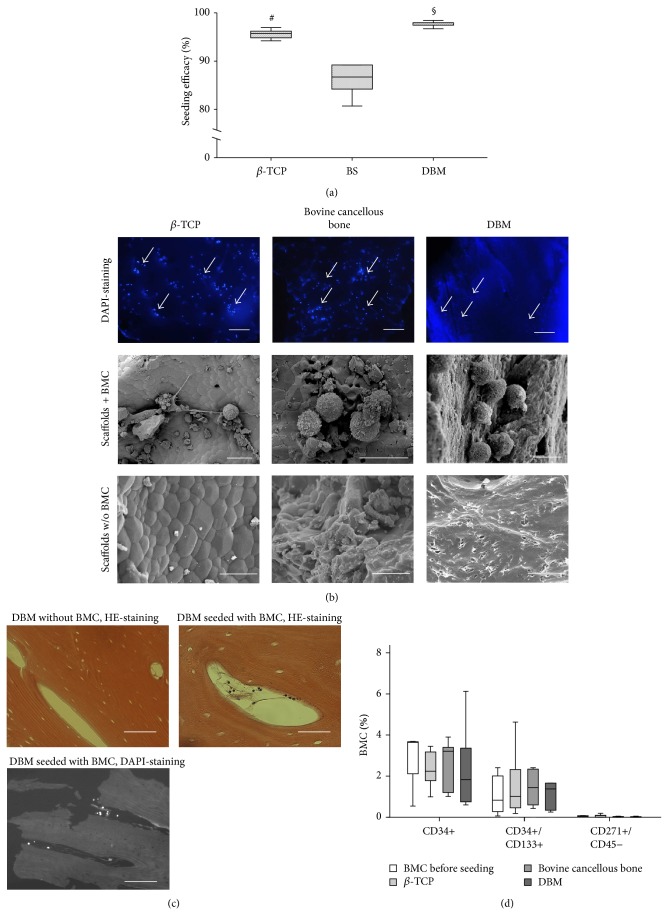
Impaired adhesion of BMC on bovine cancellous bone. (a) BMCs were seeded on uncoated β-TCP, DBM, and bovine cancellous bone (BS) and seeding efficacy was assessed by DAPI staining as described in Materials and Methods section (n = 7). § P < 0.05, DBM versus β-TCP, bovine cancellous bone; # P < 0.05 β-TCP versus bovine cancellous bone. Direct confirmation of the presence of BMC on the biomaterials is presented in (b). Upper row column shows DAPI stained BMC on the biomaterials; the middle row shows BMC on the materials' surface by SEM imaging. Cell free biomaterials were shown for comparison in the lower row. (c) BMCs were not only located on the surface of DBM but also found frequently located within the DBM as demonstrated by means of histology. (d) The percentages of progenitor cell types which resemble EPC (CD34+/CD45+; CD34+/CD133+/CD45+) and MSC (CD45−/CD271+) in the fraction of nonadhering cells were presented. No selective enrichment or depletion of progenitor cell subpopulation depending on the biomaterial were noted (n = 5). Size bars (b): 200 μm (DAPI-staining), 6 μm (SEM images, middle row), and 10 μm (SEM images lower row); space bars (c): 200 μm (up left), 400 μm (low left), and 50 μm (up right).

