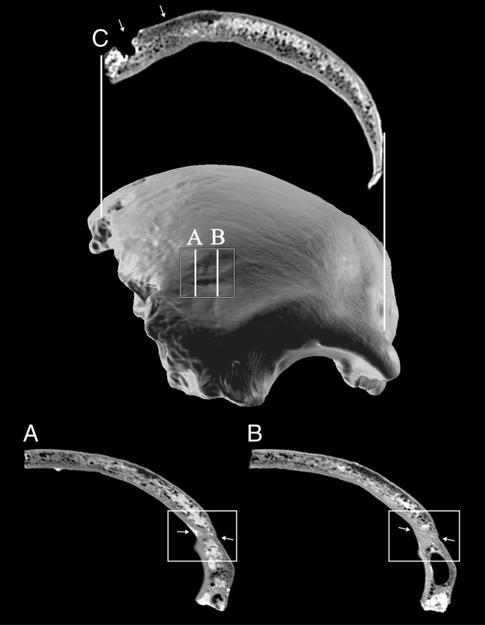
Fig. 2.
Three-dimensional model of the frontal bone in anterior view, showing the healed frontal trauma and the inner structure of the frontal squama. The zone of trauma is demarcated by the white rectangle. (A and B) Two sagittal sections intersect the healed trauma, as shown by white arrows. (C) Coronal section is given at the level of the postmortem perforation of the right side of the frontal squama (left arrow). The right arrow outlines the pathological inner structure changes of the diploe and outer table.

