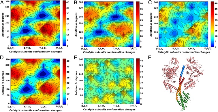
Fig. 3.
The CG electrostatic free energy surfaces for the γ-truncation systems without the addition of chemical free energy are shown for (A) γ1, (B) γ2, (C) γ3, (D) γ4, and (E) combined γ2–γ3. (F) The γ-subunit with a pair of opposing β-subunits are shown [Protein Data Bank (PDB) ID: 1H8E], where γ1, γ2, γ3, and γ4 are colored in blue, red, orange, and green, respectively.

