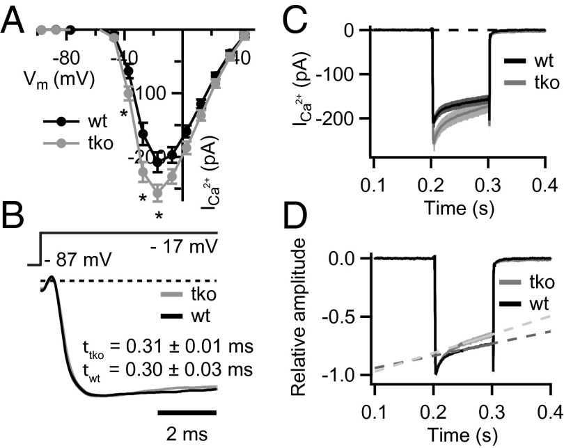
Fig. 2.
Disruption of PVα, CB, and CR enhances Ca2+-current amplitude and inactivation. (A) Current–voltage relationship of the voltage-gated Ca2+ current in TKO (gray, n = 20) and WT (black, n = 23) IHCs from 2–3-wk-old mice. Note the slightly larger Ca2+ currents in the TKO IHCs (*P = 0.01–0.03, Student t test). (B) Normalized average Ca2+ currents in response to 10–100-ms depolarizations to the peak Ca2+-current potential on an expanded time scale demonstrate that the kinetics of the activation were not different among TKO and WT IHCs (P = 0.13, Wilcoxon rank-sum test). Data were fitted with I(t) = I0 + Imax × (1 − e−t/τ)p, whereby the power (p) was fixed to 2 in most cases. (C and D) Absolute (C) and normalized (D) Ca2+ currents in response to 100-ms depolarizations to the peak Ca2+ current potential. A stronger Ca2+-current inactivation was observed in the TKO IHCs. Slopes of the linear fits (1/s) are 1.0 ± 0.1 and 1.6 ± 0.2 for the Ca2+ currents in WT (n = 16) and TKO (n = 14) IHCs, respectively (P = 0.02, Student t test).

