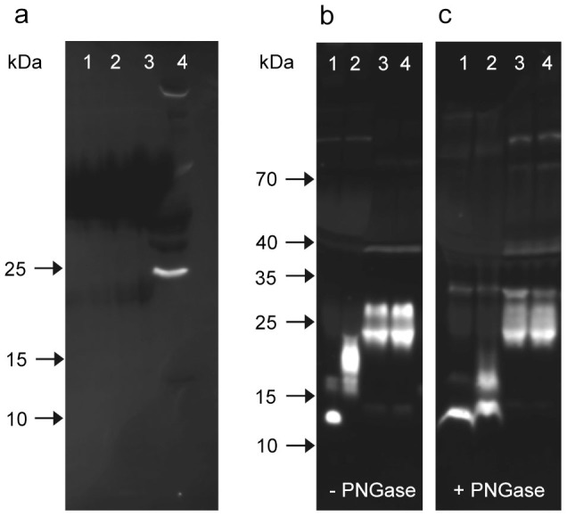
Figure 2. Western blots for detection of FNDC5 and irisin.

(a) pAb-E, which reacts with a C-terminal peptide (outside the irisin domain) was used to detect full-length FNDC5. This pAb-E detected no band in human serum samples (lanes 1–3), but mouse muscle extract showed a sharp band at ~25 kDa.(lane 4). (b, c) pAb-C was used to stain rNG-irisin (lane 1), G-irisin (lane 2) and human serum samples (lanes 3, 4) before (b) and after (c) deglycosylation with PNGase. All images were taken after 10 min of exposure without contrast enhancement.
