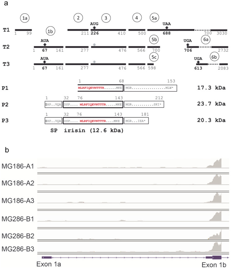
Figure 6. FNDC5 transcripts and deduced peptides.
(a) Transcript structure of human FNDC5 (T1: NM_001171941.2, T2: NM_153756.2, T3: NM_001171940.1) and deduced peptide structure (P1–P3). Numbers refer to nucleotides (T1-3) or amino acids (P1–3). Black bars represent exons. Exon numbers are given in circles. Start and stop codons are indicated above the bars. The irisin peptide is marked by a bold box. The open box marks the truncated irisin peptide in P1 theoretically resulting from transcript T1. The size of irisin and FNDC5 protein variants is given. The peptide signature identified by mass spectrometry is marked in red. SP: signal peptide. (b) Example for detection of FNDC5 transcripts by RNA-sequencing of skeletal muscle biopsies. Exon 1a is specific for transcript T1 whereas exon 1b is part of transcripts T2 and T3. The panels represent results for one individual before (A1–A3) and after 12 weeks of training intervention (B1–B3). A1 and B1 were measured before, A2 and B2 immediately after, and A3 and B3 2 hours after acute exercise13.

