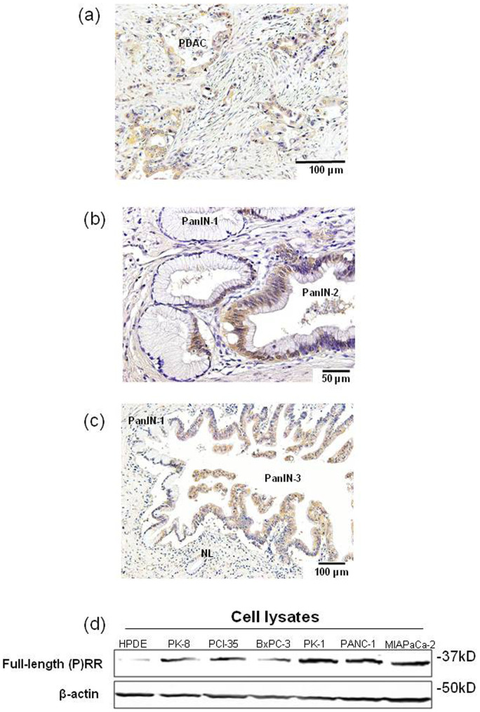
Figure 2. (P)RR is highly expressed in precursor and PDAC lesions and human PDAC cell lines.
(a) Typical immunohistochemical labeling profiles of (P)RR in PDAC tissues. (b) (P)RR expression in PanIN-1 and PanIN-2 lesions in representative pancreatic tissue samples. The PanIN-2 lesions show strong (P)RR immunoreactivity in the cytoplasm, although the PanIN-1 lesions show only focal and faint (P)RR staining. (c) (P)RR expression in normal pancreatic duct (NL), and PanIN-1 and PanIN-3 lesions in representative pancreatic tissue samples. The PanIN-3 lesions show strong (P)RR immunoreactivity in the cytoplasm. (d) Protein expression of full-length (P)RR in cell lysates was measured in HPDE cells and six different PDAC cell lines. β-actin was used as a loading control. Consistent results were observed when three experiments were repeated.

