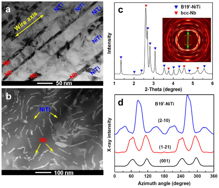
Figure 1. Typical microstructure of in-situ Nb nanowires -oriented martensitic NiTi matrix composite wire.
(a–b), Scanning transmission electron microscopic (STEM) image of the longitudinal-section and cross-section of the composite wire (bright regions: cross sections of Nb nanowires; dark regions: NiTi matrix). (c), 1D high-energy X-ray diffraction (HE-XRD) pattern. Inset is its corresponding 2D HE-XRD pattern. Green arrow represents the wire axial direction. (d), Evolution of HE-XRD intensity for multiple planes of B19′-NiTi phase along the Debye-Scherrer rings recorded on area detector diffraction image (inset of Figure 1c).

