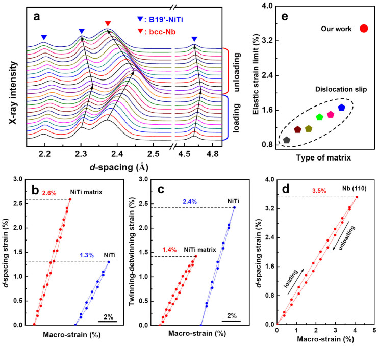
Figure 4. In situ synchrotron X-ray diffraction analysis of the NiTi-Nb composite and the binary NiTi alloy (with oriented martensite).
(a), Evolution of in situ synchrotron X-ray diffraction patterns of the NiTi-Nb composite during a tensile deformation cycle to 4% macroscopic strain. (b), The d-spacing strain with respect to applied macroscopic strain for the B19′-NiTi (001) planes perpendicular to the loading direction in the NiTi matrix of the NiTi-Nb composite (the red curve) and in the binary NiTi alloy (the blue curve). (c), The calculated twinning-detwinning strains versus the applied macroscopic strain (the red curve for the NiTi matrix in the NiTi-Nb composite; the blue curve for the binary NiTi alloy). (d), The d-spacing strain with respect to applied macroscopic strain for the Nb (110) planes perpendicular to the loading direction in the NiTi-Nb composite. (e), Comparison of the elastic strain limits of the Nb nanowires embedded in oriented martensitic NiTi matrixand embedded in the conventional metal matrix deforming by dislocation slip23,28,29,30.

