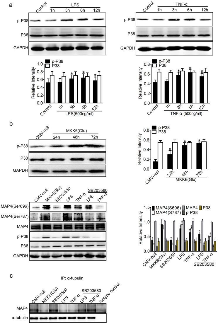
Figure 4. P38/MAPK activation mediates MAP4 phosphorylation in inflammation-induced ALI.
(a) Western blotting was used to detect phospho-p38 (p-P38) and P38 following treatment with LPS or TNF-α (500 ng/ml for 1, 3, 6, and 12 hr). *P < 0.05 vs. the control group; #P < 0.05 vs. the LPS/TNF-α (6 h) group. (b) Confirmation of MKK6 (Glu) transfection at comparable levels in HPMECs. Cells were transfected with CMV-null or MKK6 (Glu) and pretreated with SB203580 (5 μM) before the LPS or TNF-α treatment. The Western blot shows the phosphorylation of P38 and MAP4 at S696 and S787 and the total levels of MAP4 and P38. The graph shows the mean ± SEM (n = 3). *P < 0.05 vs. the CMV-null group; #P < 0.05 vs. the LPS or TNF-α group. (c) Determination of MAP4 binding to tubulin in cells with or without MKK6 (Glu) overexpression or SB203580 pretreatment under LPS or TNF-α treatment by IP; Isotype control is as the negative control (n = 3).

