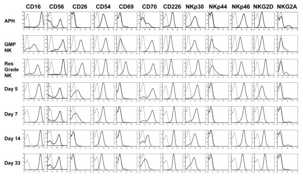
Figure 4. Immunophenotype of NK cells pre-expansion, post-expansion, and post-infusion (subject 7).
CD56 histogram overlays are gated on lymphocytes, all others are gated on CD3−CD56+ lymphocytes. Thin lines are isotype controls and thick lines are the indicated marker. The samples shown are the apheresis product analyzed the day of collection (Aph), the clinical product post-shipping (GMP NK), small scale research grade NK generated at UAMS (Res Grade NK) and PB samples obtained at the post-infusion time points indicated.

