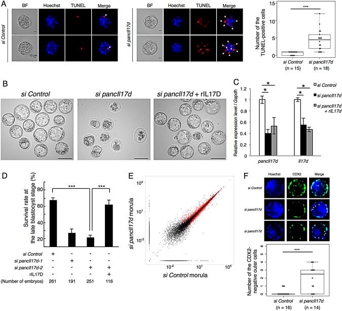
Fig. 4.
Developmental defects induced by pancIl17d knockdown and rescue by addition of recombinant IL17D protein. (A) TUNEL assay of morula embryos. Arrowheads indicate TUNEL-positive blastomeres. pancIl17d knockdown embryos showed increased TUNEL-positive cells. Two representative blastomeres are shown. To the right is a box plot of the number of TUNEL-positive cells in each embryo. (B) Morphology of late blastocysts. (C) Expression levels of pancIl17d and Il17d measured by qPCR in control, pancIl17d knockdown and rIL17D-supplemented pancIl17d knockdown morula embryos. (D) Survival rate of control and pancIl17d knockdown embryos at day 4.5 of in vitro culture. Two different siRNAs targeting pancIl17d were used. (E) Scatter plots of gene expression in control and pancIl17d knockdown morula embryos based on the RPKMs of RefSeq genes. Red dots indicate the genes that show statistically significant changes. (F) Immunostaining of CDX2 protein in control and pancIl17d knockdown late blastocyst. Arrowheads indicate CDX2-negative outer cells Beneath is a box plot of the number of CDX2-negative outer cells in each embryo *P<0.05; ***P<0.001. Error bars indicate s.e.m.

