
Figure 8.
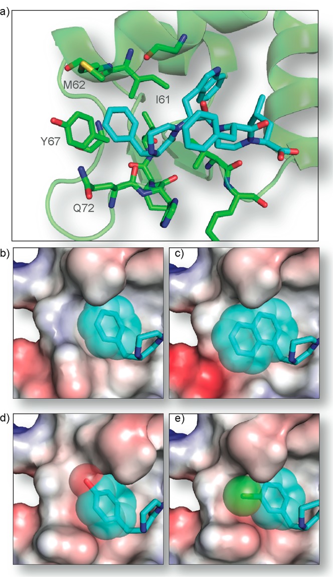
Examination of the N-terminal residue-binding pocket in Mdm2. (a) The phenylalanine residue at the R1 position of 4 (cyan) resides in a flexible pocket consisting of Ile-61, Met62, Tyr67, and Gln72 of Mdm2 (green). (b) Predicted orientations of phenylalanine and analogues (c) naphthylalanine of mimic 15, (d) tyrosine of mimic 16, and (e) 3-chlorophenylalanine of mimic 18. Electrostatic surface of Mdm2 is modeled by Pymol.