
Figure 5.
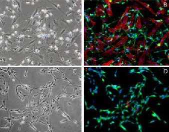
Double-immunofluorescent staining of Schwann cells with P75NTR/Thy1.1 and S100/Thy1.1 after the second round of purification. Scale bar: 40 μm.
All cells in a bipolar or tripolar shape were positive for both S100 (green) and P75NTR (green), while flat-shaped fibroblasts were Thy1-positive only (red). Nuclei were visualized by 4′-6-Diamidino-2-phenylindole staining (blue).
(A) Bright field of cells before purification (light microscope).
(B) Fluorescent view of P75NTR/Thy1.1 double staining before purification (fluorescent microscope).
(C) Bright field of cells after two rounds of purification with dispase (light microscope).
(D) Fluorescent view of S100/Thy1.1 double staining after two rounds of purification with dispase (fluorescent microscope).