Figure 2.

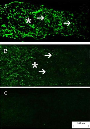
Axonal regeneration in retinal ganglion cells after optic nerve crush (immunofluorescence staining; fluorescein isothiocyanate-labeling; scale bar: 100 μm).
Representative photomicrographs show the crush site (asterisk) and regenerating axons with growth associated protein-43 immunofluorescence (arrowheads) on day 15 after injury.
Numerous growth associated protein-43 positive axons are seen passing through the crush site and they grew beyond the distal crush site in the EPO group (A). In contrast, very few axons traversed the crush site in the PBS group (B). In addition, the sham-surgery group (C) showed no staining. PBS: Phosphate buffered saline; EPO: erythropoietin.
