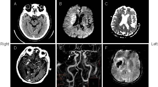
Figure 1.

A 73-year-old woman with left hemiparesis and aphasia was treated with intravenous tissue plasminogen activator within 3 hours after onset (patient 2).
(A) The admission computed tomography (CT) showed no evidence of abnormality. Initial diffusion weighted imaging (B) and apparent diffusion coefficient (C; 2.5 hours after admission CT) showed restricted diffusion in the right middle cerebral artery territory.
(D) Gd-enhanced T1-weighted image showed the hyperintense middle cerebral artery sign (arrow) in the right middle cerebral artery stem.
(E) Magnetic resonance angiography showed right middle cerebral artery stem occlusion (arrow).
(F) Follow-up of gradient echo interleaved Echo Planar Imaging on day 2 showed hemorrhage in the right middle cerebral artery territory.
