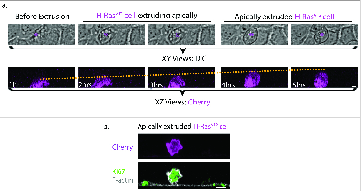
Figure 1.

Morphological comparisons between H-RasV12 expressing cell undergoing extrusion and H-RasV12 expressing cell that has been extruded. (a) Still images of brightfield (top panel, magenta asterisk indicates H-RasV12 expressing cell) and confocal fluorescence XZ cross-sections (bottom panel) from live imaging of single H-RasV12 expressing Caco-2 cells within a wild-type epithelia monolayer. The yellow dotted line in the fluorescence XZ cross-sections indicates the gradual increase in the height of the single H-RasV12 expressing cell as extrusion takes place. Based on the relative height of the extruding cell to its neighboring cell, the process of extrusion can be classified into 3 stages: Before extrusion, H-RasV12 cell extruding apically and apically extruded H-RasV12 expressing cell. (b) Immunofluorescence XZ cross-section of an apically extruded H-RasV12 expressing cell that is loosely attached on the untransfected epithelia monolayer and stained for the proliferation marker Ki67. Scale Bar: 5 μm.
