Figure 3.

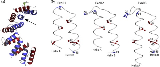
(a) Ribbon representation of the S. meliloti Rm1021 ExoRm model (residues 31–268). The repeats are numbered from 1 to 6 and helix A and helix B of each repeat are colored blue and red, respectively. The N-terminal 310 helix is colored green. The cleavage site is indicated with an arrow. (b) Backbone trace of the first three repeats of ExoR. Conserved structural residues responsible for tight packing of helices are colored red and those responsible for sharp turns in loop regions are colored blue. The numbers represent the positions of the amino acid residues in repeats.
