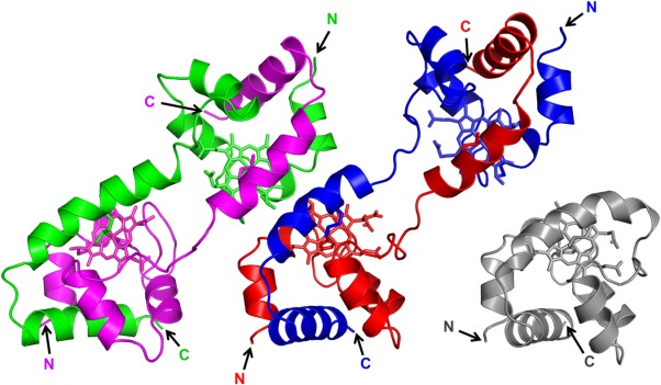
Figure 1.

Crystal structures of dimeric AA cyt c555 (PDB code: 3X15). Two dimer structures (red-blue and magenta-green) were observed in the asymmetric unit. The red and blue regions and the magenta and green regions in the dimers belong to different protomers. The hemes are shown as stick models. Side chain atoms of heme-binding Cys12 and Cys15, and heme iron-coordinating His16 and Met61 are also shown as stick models. The N- and C-termini are labeled as N, C, respectively. Structure of monomeric AA cyt c555 (gray, PDB code: 2ZXY) is also shown.
