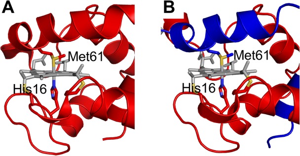
Figure 3.

Active site structures of monomeric and dimeric AA cyt c555. A) Structure of monomeric AA cyt c555 (PDB code: 2ZXY). B) Structure of dimeric AA cyt c555 solved in this study (PDB code: 3X15). The red and blue regions in the dimer belong to different protomers. Side chain atoms of His16 and Met61 are shown as stick models. The hemes are shown as gray stick models. The sulfur atoms of the side chains of Cys12, Cys15, and Met81 are depicted in yellow, and the nitrogen atoms of the side chain of His16 are depicted in blue.
