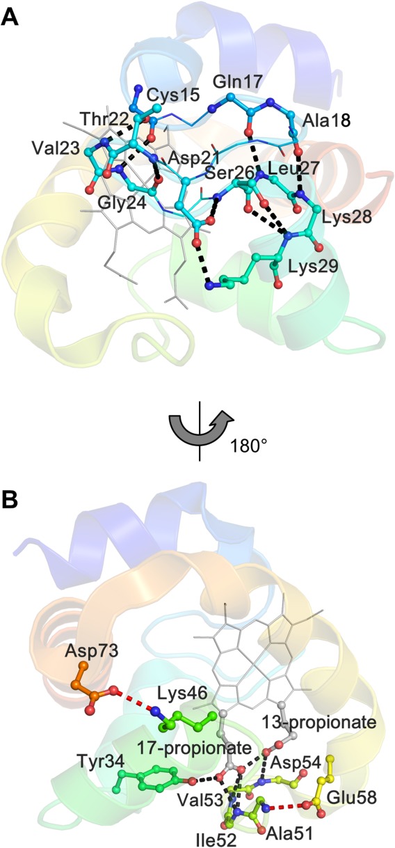
Figure 7.

Hydrogen bonds in monomeric AA cyt c555. A) Hydrogen bonds formed at Cys15−Lys29. B) Hydrogen-bonding network around the heme propionates (black), together with the hydrogen bonds formed by the extra 310-α-310 helix and the C-terminal helix, respectively, with the rest of the protein (red). The protein is depicted with sequential colors from blue for the N terminal to red for the C terminal, and the heme is depicted in gray. The main chain or side chain of the residues forming the hydrogen bonds are shown as ball and stick models, and its nitrogen and oxygen atoms are depicted in blue and red, respectively.
