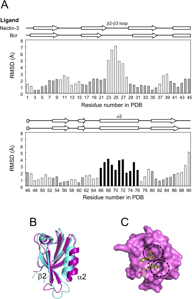
Figure 2.

Plasticity of the ligand-binding groove. (A) Backbone structural comparison. The RMSD of α-carbons in the AFPDZ bound by ligand peptides was calculated using MOLMOL.49 Amino acids comprising secondary structures in the AFPDZ–nectin-3 complex are gray in color. The most different element, α2, is highlighted in black. (B) Superposition of the AFPDZ in the AFPDZ–nectin-3 (magenta) and Bcr (light blue) complexes. A ligand-binding pocket is formed between β2 and α2. (C) Surface representation of the AFPDZ–nectin-3 complex. Binding pockets are separately formed for Val(0) and Trp(−2) [dotted circles].
