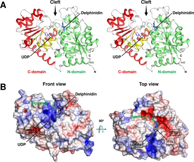
Figure 2.
(A) Stereo view of the UDP-bound form of UGT78K6. The N-domain (green) and the C-domain (red) are shown with the secondary structures. The PSPG motif comprising residues 325–368 is in yellow. The UDP molecule (light blue carbon) observed in this study is shown as a stick model. The delphinidin molecule was modeled at the acceptor-binding site of the UDP-bound form using the coordinates of the structure in complex with delphinidin (marine blue carbon). Amino acid residues discussed in the text are indicated. (An interactive view is available in the electronic version of the article.) (B) Electrostatic surface potential of UGT78K6. The electrostatic potentials of the protein surface were calculated using the APBS program24 and colored by electrostatic potential isocontours from the potential of +5 kT e−1 (blue) to −5 kT e−1 (red). Front view, same direction as Figure 2(A); top view, rotated ∼90° around the horizontal axis.

