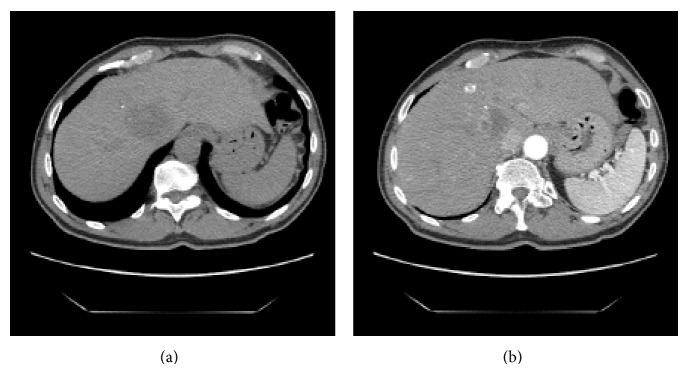
Figure 1.

Pretreatment CT scan (a) CT image showing the main tumor in the right lobe of the liver at the confluence of the right and middle hepatic veins measuring 42 × 35 mm. (b) CT-image in the arterial phase showed the second lesion in segment 4 measuring 12 mm in diameter.
