Figure 2.

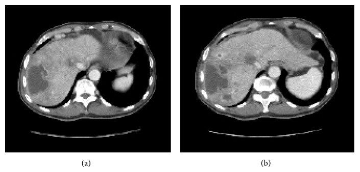
CT image at 28 d after TACE with DC beads. (a) CT image showing a huge abscess in the right lobe of the liver. (b) Many small hypodense areas are located mostly in segments 4, 6, 7, and 8 of the liver.

CT image at 28 d after TACE with DC beads. (a) CT image showing a huge abscess in the right lobe of the liver. (b) Many small hypodense areas are located mostly in segments 4, 6, 7, and 8 of the liver.