
Figure 4.
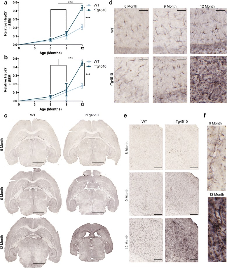
Hsp27 abundance increases with age in rTg4510 mouse brain. Hsp27 staining was quantified in the a hippocampus (± SEM) and b frontal cortex (± SEM) of 6-, 9-, and 12-month old rTg4510 and wild-type mice. ***p <0.001. Representative images of the c whole brain, d hippocampus (from CA1 region), and e frontal cortex are shown. Scale bars represent 2000 μm, 50 μm, and 200 μm respectively. High magnification images of f blood vessels in the hippocampus from 6- and 12-month old rTg4510 mice. Scale bar represents 20 μm.